An approach to the diagnosis of lumbar disc herniation using deep learning models
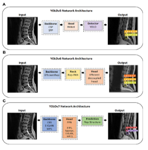
This study addresses the challenge of detecting lumbar disc herniation (LDH) in MRI due to variations in disc shape and position. Deep learning, specifically the YOLO model series (YOLOv5, YOLOv6, YOLOv7), was used to automate LDH detection. MRI images were converted, labeled, and augmented for training. Results showed that YOLOv5x with an augmented dataset achieved the highest performance, with 89.3% mAP. Non-augmented YOLOv5x also showed balanced LDH detection across lumbar regions. YOLOv5x’s strong performance suggests it can assist clinicians in early LDH diagnosis, improving patient outcomes by facilitating timely intervention.
https://www.frontiersin.org/journals/bioengineering-and-biotechnology/articles/10.3389/fbioe.2023.1247112/full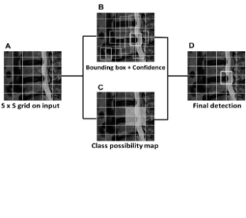
Lumbar Disc Herniation Automatic Detection in Magnetic Resonance Imaging Based on Deep Learning
Lumbar disc herniation (LDH) is a leading cause of lower back pain, often assessed through MRI, which captures spinal tissue abnormalities. This study trained a YOLOv3 deep learning model to detect LDH on a small-scale MRI dataset enhanced through data augmentation. Participants were adults with at least six months of work experience. Results showed a 92.4% mean average precision (mAP) at 550 images with augmentation, demonstrating effective detection. Data augmentation was essential in preventing model overfitting and enhancing accuracy, supporting the potential of deep learning for fast, automatic LDH screening even with limited clinical data.
https://www.frontiersin.org/journals/bioengineering-and-biotechnology/articles/10.3389/fbioe.2021.708137/full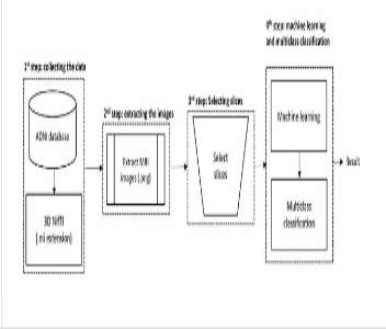
Deep Learning Applications in MRI-Based Detection of the Hippocampal Region for Alzheimer’s Diagnosis.
The hippocampal region is one of the most affected brain areas observed as a landmark in Magnetic Resonance Imaging (MRI) images for Alzheimer’s disease (AD) diagnosis. The diminished alterations in the hippocampal and degeneration of cholinergic circuits have been conclusively correlated with a decline in memory and cognitive function. However, the hippocampal region may not appear as clearly defined as other brain regions, making it difficult for neurologists and researchers to identify by visual inspection. The application of deep learning models to pinpoint the hippocampal region was initially valued. We assessed the ability of a deep learning model, You Only Live Once (YOLO), to detect hippocampal regions in three MRI image views and categories. The Alzheimer’s Disease Neuroimaging Initiative-first (ADNI−1) dataset was used with 220 subjects in three categories using the three YOLO models. We obtained the YOLO performance for hippocampal region detection with accuracy in three views and categories. The average mean Average Precision (mAP) performance accuracy for YOLOv3 was 0.87, YOLOv4 was 0.85, and YOLOv5 was 0.96, respectively. The high accuracy of the detection of the hippocampal region was remarkable. We found that the sagittal view was higher than the axial and coronal views. Simultaneously, the Mild Cognitive Impairment (MCI) in the coronal view was lower among the three models. The results showed that YOLOv5 is a suitable model for detecting the hippocampal region in MRI images, and the sagittal view is the most reliable for detecting the hippocampal region in diagnosing AD. Our findings demonstrate the importance of detecting the hippocampal region to diagnose AD and accurately analyzing the hippocampal area within the region. The YOLOv5 model substantially affected performance metrics and interpretability across the three views and categories.
https://ieeexplore.ieee.org/abstract/document/10613028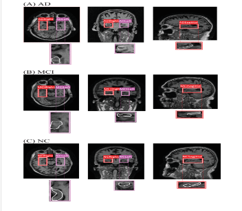
Diagnosis of Alzheimer’s disease using convolutional neural network with select slices by landmark on Hippocampus in MRI images.
Alzheimer’s disease (AD) is a major public health priority. Hippocampus is one of the most affected areas of the brain and is easily accessible as a biomarker using MRI images in machine learning for diagnosing AD. In machine learning, using entire MRI image slices showed lower accuracy for AD classification. We present the select slices method by landmarks on the hippocampus region in MRI images. This study aims to see which views of MRI images have higher accuracy for AD classification. Then, to get the value of three views and categories, we used multiclass classification with the publicly available Alzheimer’s Disease Neuroimaging Initiative (ADNI) dataset using Resnet50 and LeNet. The models were used in a total dataset of 4,500 MRI slices in three views and categories. Our study demonstrated that the selecting slices performed better than using entire slices in MRI images for AD classification. Our method improves the accuracy of machine learning, and the coronal view showed higher accuracy. This method played a significant role in improving the accuracy of machine learning performance. The results for the coronal view were similar to the medical experts usually used to diagnose AD. We also found that LeNet models became the potential model for AD classification.
https://ieeexplore.ieee.org/abstract/document/10147819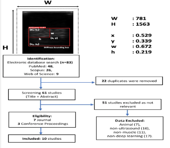
Plantar Soft Tissue Stiffness Automatic Estimation in Ultrasound Imaging using Deep learning.
Preventing diabetic foot ulcers (DFU) is critical for diabetes mellitus (DM) patients. Increased stiffness of plantar foot may cause higher plantar pressure leading to a higher risk of DFU. Soft tissue stiffness can be determined by measuring the soft tissue thickness with indentation depth and stress. Therefore, we hypothesized that the deep learning model could detect the ultrasound image pixel change under soft tissue compression. This study aimed to apply the deep learning model to analyze the ultrasound image pixel thickness of plantar foot, then predict the soft tissue indentation depth and loading force for estimating the stiffness. This study has developed a motor-driven ultrasound indentation system to apply programmable compression and simultaneously assess soft tissue mechanical properties and responses in indentation depth and loading force. In addition, the effective Young's modulus was calculated to characterize mechanical properties of soft tissues in the first metatarsal head. The deep learning method employed the YOLOv5x model to train and detect the small object in the indentation depth, such as ultrasound image pixel changes. Finally, the dataset images were processed with labeling annotation from the soft tissue indentation depth and loading force. The deep learning results showed 0.995 in mean Average Precision (mAP), 0.999 in precision, 1.000 in Recall, and 0.013 in Loss. A significant correlation was found between the ultrasound image pixel changes and soft tissue indentation depth (r = 0.98, p < 0.05). Furthermore, a significant correlation was observed between the ultrasound image pixel changes and the loading force in the first metatarsal head (r=0.85, p < 0.05). The validation and prediction models were lower than the training models in the effective Young's modulus results. However, the results of the initial modulus were similar between the three models. Our findings recommend that applying deep learning in the ultrasound image can predict soft tissue indentation depth and loading force to calculate the stiffness of the plantar foot.
https://experts.illinois.edu/en/publications/plantar-soft-tissue-stiffness-automatic-estimation-in-ultrasound-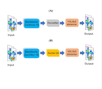
Deep learning in left and right footprint image detection based on plantar pressure.
People with cerebral palsy (CP) suffer primarily from lower-limb impairments. These impairments contribute to the abnormal performance of functional activities and ambulation. Footprints, such as plantar pressure images, are usually used to assess functional performance in people with spastic CP. Detecting left and right feet based on footprints in people with CP is a challenge due to abnormal foot progression angle and abnormal footprint patterns. Identifying left and right foot profiles in people with CP is essential to provide information on the foot orthosis, walking problems, index gait patterns, and determination of the dominant limb. Deep learning with object detection can localize and classify the object more precisely on the abnormal foot progression angle and complex footprints associated with spastic CP. This study proposes a new object detection model to auto-determine left and right footprints. The footprint images successfully represented the left and right feet with high accuracy in object detection. YOLOv4 more successfully detected the left and right feet using footprint images compared to other object detection models. YOLOv4 reached over 99.00% in various metric performances. Furthermore, detection of the right foot (majority of people’s dominant leg) was more accurate than that of the left foot (majority of people’s non-dominant leg) in different object detection models.
https://www.mdpi.com/2076-3417/12/17/8885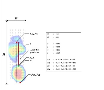
A deep learning method for foot progression angle detection in plantar pressure images.
Foot progression angle (FPA) analysis is one of the core methods to detect gait pathologies as basic information to prevent foot injury from excessive in-toeing and out-toeing. Deep learning-based object detection can assist in measuring the FPA through plantar pressure images. This study aims to establish a precision model for determining the FPA. The precision detection of FPA can provide information with in-toeing, out-toeing, and rearfoot kinematics to evaluate the effect of physical therapy programs on knee pain and knee osteoarthritis. We analyzed a total of 1424 plantar images with three different You Only Look Once (YOLO) networks: YOLO v3, v4, and v5x, to obtain a suitable model for FPA detection. YOLOv4 showed higher performance of the profile-box, with average precision in the left foot of 100.00% and the right foot of 99.78%, respectively. Besides, in detecting the foot angle-box, the ground-truth has similar results with YOLOv4 (5.58 ± 0.10° vs. 5.86 ± 0.09°, p = 0.013). In contrast, there was a significant difference in FPA between ground-truth vs. YOLOv3 (5.58 ± 0.10° vs. 6.07 ± 0.06°, p < 0.001), and ground-truth vs. YOLOv5x (5.58 ± 0.10° vs. 6.75 ± 0.06°, p < 0.001). This result implies that deep learning with YOLOv4 can enhance the detection of FPA.
https://www.mdpi.com/1424-8220/22/7/2786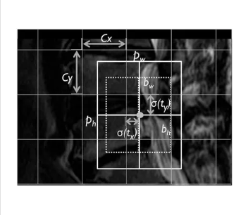
A Review of the Challenges in Deep Learning for Skeletal and Smooth Muscle Ultrasound Images
Deep learning has aided in the improvement of diagnosis identification, evaluation, and the interpretation of muscle ultrasound images, which may benefit clinical personnel. Muscle ultrasound images presents challenges such as low image quality due to noise, insufficient data, and different characteristics between skeletal and smooth muscles that can affect the effectiveness of deep learning results. From 2018 to 2020, deep learning has the improved solutions used to overcome these challenges; however, deep learning solutions for ultrasound images have not been compared to the conditions and strategies used to comprehend the current state of knowledge for handling skeletal and smooth muscle ultrasound images. This study aims to look at the challenges and trends of deep learning performance, especially in regard to overcoming muscle ultrasound image problems such as low image quality, muscle movement in skeletal muscles, and muscle thickness in smooth muscles. Skeletal muscle segmentation presents difficulties due to the regular movement of muscles and resulting noise, recording data through skipped connections, and modified layers required for upsampling. In skeletal muscle classification, the problems faced are area-specific, thus making a cropping strategy useful. Furthermore, there is no need to add additional layer modifications for smooth muscle segmentation as muscle thickness is the main problem in such cases.
https://www.mdpi.com/2076-3417/11/9/4021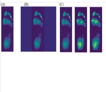
Using deep learning methods to predict walking intensity from plantar pressure images.
People with diabetes are recommended to perform exercise such as brisk walking to maintain their health. However, a fast walking speed can increase plantar pressure, especially at the forefoot and rearfoot areas, thereby increasing the risk of diabetic foot ulcers (DFU). The deep learning model can identify plantar pressure patterns for an early detection of DFU when performing various intensities of exercise. Therefore, this study aimed to identify differences in walking speeds to the plantar pressure response using deep learning methods, including Resnet50, InceptionV3, and MobileNets. The deep learning models were used to classify the plantar pressure images of healthy people walking on a treadmill. The design consisted of three walking speeds (1.8 mph, 3.6 mph, and 5.4 mph). Through 5-fold cross-validation, accuracy, and robustness, the Resnet50 model had a better performance compared to the other two models in the image classification with a mean F1 score of 0.8646 and a standard deviation of 0.0466. The results indicated that the Resnet50 model can be used to analyze plantar pressure images for assessing risks of DFU.
https://link.springer.com/chapter/10.1007/978-3-030-80713-9_35